Carbotrace

Carbotrace 680 is our red optotracer for labeling repetitive motifs in proteins and carbohydrates. Generally, Carbotrace labeling is strongest with protein aggregates. Homoglucans like cellulose are labeled reliably, albeit with lower quantum yield. Heteroglucans with low degree of branching like starches are labeled and can be distingiushed by their spectral fingerpint. Generally labelling of hemicelluloses is weak or absent.
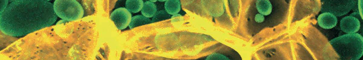
Specifically, Carbotrace 680 has been used to image cellulose and starch components in plant cells and differentiate between those components using multi-laser/multi-detector analysis with confocal microscopy. Using a fluorescence spectrophotometer to acquire excitation and emission spectra, a unique spectral signature allows to differentiate between α(1-3), α(1-4), α(1-6), β(1-3), β(1-4) and β(1-6) linked glucans. As such, Carbotrace 680 labels cell wall components such as Cellulose, Chitin, Laminarin and the hemicellulose xyloglucan. Further, Carbotrace 680 has been used to determine relative carbohydrate content and elasticity of cell walls in plant material. Contact us to learn more about applications for Carbotrace 680.
| Exmax | Emmax | Recommended filter-sets | |
|---|---|---|---|
| Carbotrace 680 | 530 nm | 680 nm | PI, mCherry, Cy3.5 |
Carbotrace 680 is available in four different formulations (See volumes and prices in the drop-down list below):
- Aqueous: 1 mg/ml solution in ultrapure water. The product should be diluted 1:1000 before use. To prevent evaporation of the aqueous solvent, close the container carefully after use, spin down liquid and use up small volumes quickly.
- DMSO: 1 mg/ml solution in DMSO to prevent solvent evaporation. The product should be diluted 1:1000 before use.
- Solid: 1 mg solid lyophilised in a sterile injection bottle.
- Drop&Shine: 5 ml ready-to-use product in mounting medium. Ideal for use in sections. Add a some Drop&Shine and mount your slide to analyse biomass or other cellulosic materials within minutes.
Carbotrace are optotracers for anatomical mapping of carbohydrate structures in plants and for non-destructive composition analysis of glucans in bio-based materials and biofuels.
The optotracing technology entails the use of structure-responsive fluorescent molecules (optotracers) which become fluorescent when binding to a target. The excitation and emission spectra of the optotracer molecule contain information on the structure of their binding partner and their environment. This spectral fingerprint can be used to identify biomolecules in various types of materials.
Carbotrace is available in five variants which bind to repetitive motifs in proteins and carbohydrates. It has been shown that Carbotrace 680 binds to homoglucans and heteroglucans with a limited degree of branching. By using the spectral fingerprint from the excitation- and emission spectra, it was shown that the optotracer can differentiate between different types of glycosidic bonds. Using Multi-laser/Multi-detector imaging, non-destructive composition analysis of biomass has been performed such as differentiation of cellulose cell walls and starch granules in fresh potato as well as cellulose and lignin in in various types of biomass. Using fluorescence lifetime imaging in combination with Carbotrace 680, the mechanism of paper ageing was analysed. Further, Carbotrace 680 was used to visualise cellulose content in terrestrial plants (maize root and ulvophyceae) as well as algae and to characterise a cellulose immobilisation matrix in a microfluidic device. Carbotrace 480 was used as a tool for cellulose visualisation and to optimise cellulose extraction from macroalgae.
Carbotrace variant work in a wide range of salt and pH conditions. When the pH is altered during the experiment, pH controls should be included. Carotrace can be used with fluorescence plate readers, fluorescence microscopes and confocal laser scanning microscopes and fluorescence life time imaging.
We named our Carbotrace molecules after their peak emission wavelength when they are bound to their target. That means, when Carbotrace is bound to a target, it will emit fluorescence at peak emission indicated by the number associated with its name.
To view the excitation and emission spectra, please select your Carbotrace below :
2026
-
Płachno, B. J., Kapusta, M., Feldo, M., Stolarczyk, P., & Świątek, P. (2026). Immunocytochemical Analysis of the Wall Ingrowths and Cell Wall Microdomains in the Digestive Glands of Venus’ Flytrap. International Journal of Molecular Sciences. 27(3), 1193 https://doi.org/10.3390/IJMS27031193/S1
-
Herting, G., Blomberg, E., Khort, A., Rogö, H., Palmi, K., Hammar, H., Richter-Dahlfors, A., & Odnevall, I. (2026). Mechanistic insights on surface adsorption of rice-based biomolecules on stainless steel 316L and its effects on corrosion and metal migration. Journal of Food Engineering, 413, 113018. https://doi.org/10.1016/J.JFOODENG.2026.113018
2025
-
Bogdziewiez, L., Froeling, R., Schöppl, P., Juquel, J., Antoniadi, I., Skalický, V., Mathey, A., Fattaccioli, J., Sprakel, J., & Verger, S. (2025). The Q-Warg Pipeline: A Robust and Versatile Workflow for Quantitative Analysis of Protoplast Culture Conditions. Plant Direct, 9(7), e70090. https://doi.org/10.1002/pld3.70090
-
Kapusta, M., Narajczyk, M., & Płachno, B. J. (2025). Arabinogalactan Proteins Mark the Generative Cell–Vegetative Cell Interface in Monocotyledonous Pollen Grains. Cells, Vol. 14, Page 1549, 14(19), 1549. https://doi.org/10.3390/cells14191549
-
Płachno, B. J., Kapusta, M., Feldo, M., Stolarczyk, P., Małota, K., & Banaś, K. (2025). External Glands of Nepenthes Traps: Structure and Potential Function. International Journal of Molecular Sciences, 26(16), 7788. https://doi.org/10.3390/ijms26167788
-
Costa, L., Carvalho, A. F., Fernandes, A. J. S., Campos, T., Dourado, N., Rodrigues, A. C., Sampaio, P., Costa, F. M., Dourado, F., & Gama, M. (2025). On the microstructural anisotropy and mechanical properties of bacterial nanocellulose obtained by static culture. International Journal of Biological Macromolecules, 330, 148114. https://doi.org/10.1016/j.ijbiomac.2025.148114
-
Fatima, I., Wakade, G., & Daniell, H. (2025). Expression of mannanase and glucanases in lettuce chloroplasts and functional evaluation of enzyme cocktail against Candida albicans in oral cancer patient samples. Plant Biotechnology Journal, 23(7), 2689–2703. https://doi.org/10.1111/pbi.70046
-
Mao, A., Gebhard, A. C., Ezazi, N. Z., Salhotra, A., Riazanova, A. v., Shanker, R., Wågberg, L., Nielsen, L. H., & Svagan, A. J. (2025). Plant cell–inspired colon-targeted cargo delivery systems with dual-triggered release mechanisms. Science Advances, 11(20), 2653. https://doi.org/10.1126/sciadv.adt2653
-
Rueckel, M., & Pasquali, G. A. M. (2025). Spatial mapping of xylanase activity on maize meal. Animal Feed Science and Technology, 330, 116524. https://doi.org/10.1016/j.anifeedsci.2025.116524
-
Schmidt, A. E. M., Richter-Dahlfors, A., & Edlund, U. (2025). Exploring the role of lignocellulose anatomy in the production and properties of lignin-containing microfibrillated cellulose from Lupinus angustifolius. Industrial Crops and Products, 237, 122262. https://doi.org/10.1016/j.indcrop.2025.122262
-
Schmidt, A. E. M., Steinhagen, S., Nilsson, K. P. R., Edlund, U., & Richter-Dahlfors, A. (2025). Spatial in situ mapping of cellulose and other biopolymers reveals the 3D tissue architecture in the green algae Ulva fenestrata. International Journal of Biological Macromolecules, 320, 145632. https://doi.org/10.1016/j.ijbiomac.2025.145632
-
Fatima, I., Wakade, G., Ahmad, N., & Daniell, H. (2025). Expression of endochitinase and exochitinase in lettuce chloroplasts increases plant biomass and kills fungal pathogen Candida albicans. Plant Biotechnology Journal, 23(5), 1437–1451. https://doi.org/10.1111/pbi.14596
2024
-
Płachno, B. J., Kapusta, M., Feldo, M., & Swiatek, P. (2024) Homogalacturonans and Hemicelluloses in the External Glands of Utricularia dichotoma Traps. International Journal of Molecular Sciences. https://www.mdpi.com/1422-0067/25/23/13124
-
Schmidt, A. E. M., Choong, F. X., Richter‐Dahlfors, A., & Edlund, U. (2024). Defibrillated Lignocellulose Recovery Guided by Plant Chemistry and Anatomy – A Pioneering Study with Lupinus angustifolius. Advanced Sustainable Systems. https://doi.org/10.1002/adsu.202300632
-
Ferrara, V., Vetri, V., Pignataro, B., Chillura Martino, D. F., & Sancataldo, G. (2024). Phasor-FLIM analysis of cellulose paper ageing mechanism with carbotrace 680 dye. International Journal of Biological Macromolecules , 260, 129452. https://doi.org/10.1016/j.ijbiomac.2024.129452
2023
-
Inthalaeng, N., Dugmore, T. I. J., & Matharu, A. S. (2023). Production of Hydrogels from Microwave-Assisted Hydrothermal Fractionation of Blackcurrant Pomace. Gels, 9(9). https://doi.org/10.3390/gels9090674
-
Petrova, A., Ageeva, M., & Kozlova, L. (2023). Root growth of monocotyledons and dicotyledons is limited by different tissues. Plant Journal, 116(5), 1462–1476. https://doi.org/10.1111/tpj.16440
-
Holzinger, A., Plag, N., Karsten, U., & Glaser, K. (2023). Terrestrial Trentepohlia sp. (Ulvophyceae) from alpine and coastal collection sites show strong desiccation tolerance and broad light and temperature adaptation. Protoplasma. https://doi.org/10.1007/s00709-023-01866-2
2022
-
Zitzmann, F. L., Ward, E., & Matharu, A. S. (2022). Use of Carbotrace 480 as a Probe for Cellulose and Hydrogel Formation from Defibrillated Microalgae. Gels, 8(6), 383. https://doi.org/10.3390/gels8060383
-
Petrova, A., Sibgatullina, G., Gorshkova, T., & Kozlova, L. (2022). Dynamics of cell wall polysaccharides during the elongation growth of rye primary roots. Planta, 255(5), 108. https://doi.org/10.1007/s00425-022-03887-2
2020
-
Petrova, A., Gorshkova, T., & Kozlova, L. (2020). Gradients of cell wall nano-mechanical properties along and across elongating primary roots of maize. Journal of Experimental Botany. https://doi.org/10.1093/jxb/eraa561
-
Kumar, T., Soares, R. R. G., Dholey, L. A., Ramachandraiah, H., Aval, N. A., Aljadi, Z., Pettersson, T., & Russom, A. (2020). Multi-layer assembly of cellulose nanofibrils in a microfluidic device for the selective capture and release of viable tumor cells from whole blood. Nanoscale, 42. https://doi.org/10.1039/d0nr05375a
2019
-
Choong, F. X., Lantz, L., Shirani, H., Schulz, A., Nilsson, K. P. R., Edlund, U., & Richter-Dahlfors, A. (2019). Stereochemical identification of glucans by a donor–acceptor–donor conjugated pentamer enables multi-carbohydrate anatomical mapping in plant tissues. Cellulose, 26(7), 4253–4264. https://doi.org/10.1007/s10570-019-02381-5
Terms & Conditions
For detailed information on Terms & Conditions, please click the link below.
Privacy Policy
To read more how we handle personal information, please click the link below.
Support
If you have any questions related to our products or services, please contact us.