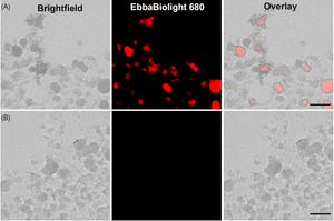
Urinary tract infection (UTI) is a common type of infection of the urinary system caused by uropathogenic Escherichia coli (UPEC). Unsurprisingly, the microenvironment of the urinary tract provides a challenge for colonisation as bacteria are exposed to hydrodynamic shear stress. To be able to understand how UPEC colonises the urinary tract, researchers from AIMES, Karolinska Institutet, Sweden developed a biomimetic proximal-tubule-on-chip (PToC) device which allowed them to monitor UPEC attachment to renal epithelial cells under the shear stress. When the researchers injected UPEC (CFT073) wild-type bacteria, single cell tracking showed that most of the bacteria just passed through the device without adhering and only a tiny portion of bacteria adhered to the epithelial cells with a few bacteria managing to strongly resist displacement by the flow and were defined as bound bacteria. Nevertheless, using wild-type CFT073, a small number of bacteria with strong adhesion were enough to initiate colony formation. To explore the molecular mechanism leading to successful attachment under shear stress the researchers created a P fimbriae (∆papG) mutant and showed that ∆papG bacteria have a reduced ability to mediate long-term adhesion under shear stress. When the researchers analysed the nature of microcolony formation in the PToC, they found that the adherent cells divide rapidly pointing out that a strong initial adhesion combined with rapid proliferation led to UPEC microcolony formation within 3–4 h. Microcolony formation was shown to depend on Type 1 Fimbriae (FimH), but not on P Fimbriae (PapG). Using EbbaBiolight, it was shown that formation of extracellular matrix (ECM) did not play a role at early time points, but was demonstrated to be established after 4 days in the biomimetic device.
Image: Detection of extracellular matrix components in UPEC microcolonies formed in a biomimetic proximal-tubule-on-chip (PToC) device with EbbaBiolight 680 after 4 days of infection. Upper panel (A) shows microcolony labelled with EbbaBiolight 680 and lower panel (B) shows controls without EbbaBiolight 680. Image from Figure 6AB by Antypas, H. et al. (2023) FEMS Microbes, 4,1–12 (CC BY).
