Labeling of surface biofilm using EbbaBiolight
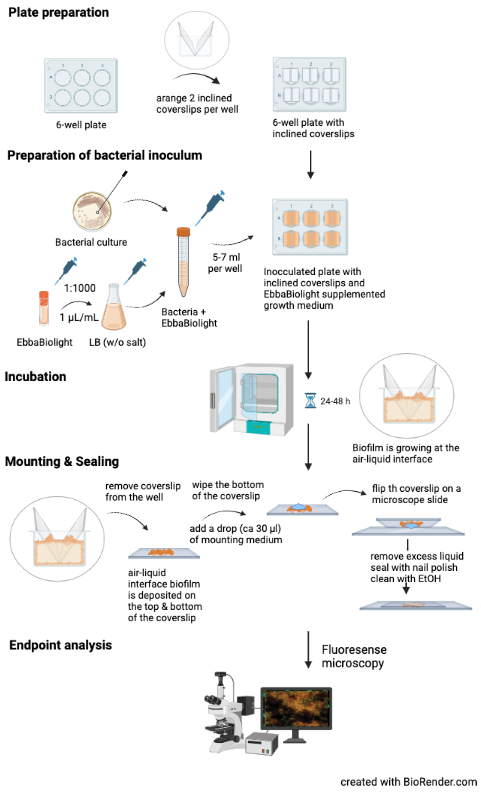
This protocol describes how to grow Salmonella biofilm at an air-liquid interface using inclined glass coverslips and how to visualize Salmonella extracellular matrix component curli using EbbaBiolight. The method described here is based on Choong et al. (2016) npj Biofilms and Microbiomes, 2, 16024 where isogenic mutants of S. Enteritidis were used to identify the extracellular matrix components curli and cellulose as targets for optotracer binding. When used as recommended, EbbaBiolight does not label Salmonella cell wall and does not influence biofilm formation. If adding EbbaBiolight during biofilm growth is not feasible, it can also be applied after the biofilm has assembled and incubated for 30-60 min.
Materials:
Equipment:
Assay Procedure:
Note: When adapting this technique, please make sure to include relevant controls to verify that EbbaBiolight does not affect biofilm formation, to confirm curli as EbbaBiolight binding target and to exclude pH effects. Please be aware that fixation might alter the staining pattern of EbbaBiolight.

Materials:
- EbbaBiolight
- LB broth (w/o salt)
- Bacteria on standard culture plate
- Sterile glass coverslips (24x24 mm)
- 6-well plate with cover or adhesive seal
- Mounting medium
- Nail polish
- EtOH 70%
Equipment:
- Incubator (28°C)
- Shaking Incubator (37°C)
- Fluorescence microscope
Assay Procedure:
- Plate preparation:
- place two sterile glass coverslips opposite to each other and inclined towards the walls of the wells in a 6-well plate.
-
Prepare bacterial inoculum:
- Pick a colony from a standard culture plate.
- Transfer colony to LB broth.
- Prepare an overnight or exponential culture under continuous shaking at 37°C.
- Dilute bacterial culture 1:100 in fresh LB broth.
- Add EbbaBiolight (1:1000) and mix gently.
- Pipett 6 ml of EbbaBiolight supplemented bacterial culture into each well fitted with glass coverslips.
- Incubation:
- Seal the plate with cover or adhesive seal & incubate at a suitable temperature for 24-48 h.
- Mounting & Sealing:
- Remove the glass coverslips from each well & wipe the backside.
- Add a drop (ca 30 µl) of mounting medium to the sample and flip the coverslip on a microscope slide.
- Remove excess liquid, seal the coverslip with nail polish & clean the slide with 70% EtOH.
- Endpoint analysis:
- Visualize biofilm using fluorescence microscopy. Use filter-sets or excitation- and emission parameters as indicated in the table below.
Optotracing with EbbaBiolight
EbbaBiolight fluorescent tracer molecules are optotracers. Unlike conventional fluorescent dyes, optotracers bind promiscuously to a range of targets with repetitive motifs. EbbaBiolight has been shown to bind to curli and cellulose in Salmonella extracellular matrix [1,2], peptidoglycan and lipoteichoic acids in the cell envelope of Staphylococci [3], β-glucans from S. cerevisiae and Chitin in C. albicans [4]. Upon binding, the fluorescence intensity of the optotracer increases. This property makes it possible to use EbbaBiolight for live fluorescent tracking of microorganisms, without the need to wash away unbound molecules. It is possible to read out fluorescence intensity at the emission maximum (Emmax) when excited at or close to the excitation maximum (Exmax). This is useful for microscopy or fluorescence spectroscopy when straight-forward data analysis is required. Yet, due to the unique properties of the optotracers, a unique optical fingerprint is produced reflecting the specific nature of the target (sample composition) and environment (pH, osmolarity, polarity of the medium). This means that depending on the specific properties of the sample, Exmax or Emmax can shift, or the appearance of double peaks or shoulders might indicate binding to multiple targets. We therefore recommend acquiring fluorescence excitation and emission spectra whenever possible within experimental limitations. EbbaBiolight excitation- and emission spectra can be accessed here.
| Exmax | Emmax | Excitation spectrum (detect at Emmax) | Emission spectrum (excite at Exmax) | Recommended filter-sets |
|
|---|---|---|---|---|---|
| EbbaBiolight 480 | 420 nm | 480 nm | 300 - 450 nm | 450 - 800 nm | DAPI |
| EbbaBiolight 520 | 460 nm | 520 nm | 300 - 490 nm | 490 - 800 nm | FITC, GFP |
| EbbaBiolight 540 | 480 nm | 540 nm | 300 - 510 nm | 510 - 800 nm | FITC, GFP, YFP |
| EbbaBiolight 630 | 520 nm | 630 nm | 300 - 600 nm | 550 - 800 nm | PI, Cy3, TxRed, mCherry, Cy3.5 |
| EbbaBiolight 680 | 530 nm | 680 nm | 300 - 650 nm | 660 - 800 nm | PI, mCherry, Cy3.5 |
Read More:
- Choong FX et al. (2016) Real-Time optotracing of curli and cellulose in live Salmonella biofilms using luminescent oligothiophenes. npj Biofilms and Microbiomes, 2, 16024
- Choong FX et al. (2021) A semi high-throughput method for real-time monitoring of curli producing Salmonella biofilms on air-solid interfaces. Biofilm, 3, 100060
- Butina K. et al. (2020) Optotracing for selective fluorescence-based detection, visualization and quantification of live S. aureus in real-time. npj Biofilms and Microbiomes, 6(1), 35
- Kärkkäinen, E. et al. (2022) Optotracing for live selective fluorescence-based detection of Candida albicans biofilms. Frontiers in Cellular and Infection Microbiology, 12, 2235-2988
