Biomaterials composed of self-assembling protein building blocks are being actively explored for biomedical applications, including drug delivery, tissue engineering scaffolds, and functionalized surface coatings. Among these, spider silk has attracted particular attention due to its biocompatibility and stability. However, large-scale production of natural spider silk remains challenging, which has led to the development of recombinant spider silk proteins such as FN-silk (FN-4RepCT), functionalized with a cell-binding motif (RGD). FN-silk has previously been shown to form fibrillar structures at air-liquid interfaces, and more recently, to self-assemble into microspheres within physiological buffer (PBS) solutions.
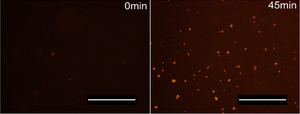
In this study, Ornithopoulou et al. (2023) used several advanced imaging and structural analysis methods to investigate the self-assembly process of FN-silk into microspheres and to analyse their properties. Using Amytracker 680 with fluorescence microscopy, the authors visualized the protein secondary structure transition during self-assembly. No fluorescence was detected before microsphere formation, but signal intensity increased within 10 min and continued over time, indicating progressive microsphere assembly and confirming the strong presence of β-sheets in the FN-silk microspheres.
Finally, they demonstrated that FN-silk microspheres can be effectively used for biofunctionalization of cell culture surfaces and can integrate into cell spheroids, highlighting their potential for targeted delivery of drugs or growth factors in future biomedical applications. Altogether, these findings enhance our understanding of silk-protein self-assembly mechanisms and support the use of silk microspheres as additives for cell culture applications.
Image: Amytracker 680 (red) visualising FN-silk microsphere formation. Imaged using Fluorescence microscopy. Scale bar: 200 μm. Adapted from Fig. 5C, Ornithopoulou et al. 2023, (CC BY 4.0).
