Monitoring heat-induced swelling of Starch granules
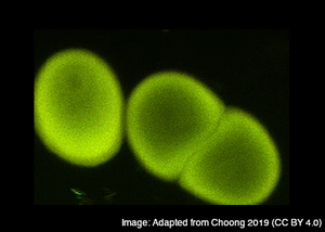
As starch is a thermo-sensitive material that undergoes drastic morphological changes when heated, it is even possible to monitor starch reorganization during heat-induced swelling. When freshly extracted potato starch granules marked with Carbotrace 680 are heated from 20 °C to 90 °C and imaged with a confocal microscope, it becomes obvious that no change in granule size and morphology occurs until the granules are heated to 60 °C. At temperatures higher than 60 °C swelling is observed which can be identified by growing starch granules, but also by a visible change in the fluorescence color and intensity produced by Carbotrace 680 (See image).
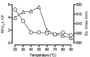
Fluorescence intensity (RFU) and color (determined by the wavelength at the excitation maximum, λ - Exmax) can be easily quantified using a fluorescence plate reader. This method greatly improves and simplifies the detection of heat-induced morphological changes in starch, since minor rearrangements in starch packing and swelling can be identified. While the RFU gives an indication on the onset of swelling, λ - Exmax identifies early, heat-induced starch rearrangement, long before morphological changes become visible.
Image: Freshly extracted potato starch granules marked with Carbotrace 680 are heated from 20 °C to 90 °C and the fluorescence intensity (RFU, circels) and color (λ - Exmax, triangles) are quantified using a fluorescence plate reader. The image is reproduced from Choong et al. 2019 (CC BY 4.0).
Detailed analysis of starch structure and morphology enabled by Carbotrace 680 can facilitate quality management of starch raw materials for use in food and pharmaceutical industries where starch reaction with water plays a pivotal role for texture and taste as well as for packing and release kinetics of pharmaceuticals.
Read More:
Carbotrace fluorescence spectra
We named our Carbotrace molecules after their peak emission wavelength when they are bound to their target. That means, when Carbotrace is bound to a target, it will emit fluorescence at peak emission indicated by the number associated with its name.
To view the excitation and emission spectra, please select your Carbotrace below :
Carbotrace 680 for monitoring starch
Detection of polysaccharides using Carbotrace 680 relies on regularly occurring units joined by glycosidic linkages. The specific type of glycosidic linkage and the occurrence of branches allow Carbotrace 680 to differentiate between different types of glucans. By means of its structure-responsive properties, Carbotrace 680 can differentiate between β(1-4) linked cellulose and α(1-4) configured amylose, but it can also detect differences between amylose and amylopectin, which contains α(1-6) linked branches.
Since starches from different sources often differ by the length of glucose chains or the amylose/amylopectin ratio, and the protein and fat content of the storage organs, Carbotrace 680 can distinguish between different source of starch like starch obtained from corn or potato.
Monitoring freshly obtained potato starch granules with Carbotrace 680 shows very fine details of the morphology of native starch granules at a sub-cellular scale. The movie shows Carbotrace 680 fluorescence when bound to potato starch granules while stepping through a stack of images collected on the confocal microscope. The most intense green fluorescence is observed on the exposed surface and the outer layer of granules, decreasing towards the core. Faint striations, likely corresponding to alternating layers in crystalline and amorphous lamina were observed in the larger granules. This pattern is likely attributed to differences in polysaccharide packing density in each lamina.
Movie: Carbotrace 680 fluorescence when bound to potato starch granules while stepping through a stack of images collected on the confocal microscope. The movie is reproduced from Choong et al. 2019 (CC BY 4.0)