Alpha-synuclein (α-Syn) aggregation hallmarks a group of neurodegenerative diseases known as synucleinopathies with Parkinson’s disease as its most well-known representative. The molecular mechanism behind the aggregation of α-Syn is still not completely understood. Many aggregation-prone proteins, like α-Syn, are known to form phase-separated condensates and recent findings suggest that the early events that lead to protein aggregation, amyloid deposition, and ultimately neurodegeneration, are happening within these condensates. Recent work from Piroska et al. published in the journal Science Advances found strong evidence of a connection between phase separation and aggregation of α-Syn. When α-Syn was present in form of a fusion protein with GFP and fragments of a faulty chaperone that facilitates multivalent interactions and formation of phase separated condensates, treatment of SH-SY5Y cells with pre-formed fibrils (PFFs) leads to formation of solid structures forming needle-like protrusions within a few hours. These α-Syn condensates show the characteristics of amyloid deposits, as they are not dissolved by detergents and stop exchanging components with the surrounding cytoplasm. Moreover, they are intensely labelled by Amytracker 630, confirming the presence of amyloid structures within them, while α-Syn condensates formed before exposure to pre-formed fibrils are not. In other words, when α-Syn separates into condensates it is more prone to aggregation and this is probably due to the elevated local concentration of α-Syn within these aggregates. Linking supersaturation of α-Syn in phase-separated compartments with amyloid formation gives important insights into the early stages of neurodegenerative conditions and might provide clues on how to interfere with preventative measures.
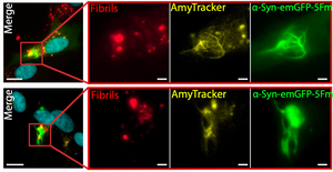
Image: Pre-formed fibrils trigger the evolution of α-Syn condensates into solid-needle-like structures in SH-SY5Y neuronal cells. Nuclei (cyan), pre-formed α-Syn fibrils (red), Amytracker (yellow), and α-Syn-GFP (green). Image from Figure S6B, Piroska et al. (2023) Science Advances, 9(33), eadg5663 (CC-BY-4.0).