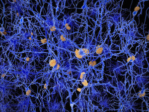
A team of researchers from Massachusetts General Hospital and Harvard Medical School as well as Linköping University have used a fluorescent probe of the same type as Amytracker for multiphoton imaging of Amyloid-β deposits in transgenic mice in vivo.
The fluorescent molecule clearly targeted and labeled core plaques in the cerebral tissue and vasculature when imaged after intravenous injection. The fluorescent signal appeared already shortly after injection, however reaching maximal intensity between 24 and 72 hours – clearly showing the capacity of the probe to pass the blood brain barrier without causing toxicity. Since core plaques are labeled intensely with the fluorescent molecule, emitting in the red channel, the molecule can also be combined with other markers for diffuse plaques or neurofibrillary tangles with emission in the green or blue channel.